Mango is not only unique in flavor but also high in nutritional value, and is one of the important tropical cultivated fruits in China. Fungal diseases are extremely important constraints in mango production, has been reported as many as 80 kinds of fungal diseases of mango, of which the most common for mango anthracnose, powdery mildew, leaf spot disease, rheumatism, etc., but also to the mango leaf spot disease occurs more common, more harmful, and a wider range of hosts. In the current agricultural production, the prevention and control of mango diseases are still dominated by highly efficient and fast chemicals, but the chemicals have adverse effects on the environment and the yield and quality of mango, therefore, it is important to carry out green prevention and control to promote the quality and healthy development of China’s mango industry.

Nanomaterials have extraordinary properties such as surface and interfacial effects, quantum size effects, small size effects and macroscopic effects, and thus have received widespread attention. Currently, nanomaterials have been applied in many fields such as environmental energy, aerospace, biomedicine and so on. Nanotechnology has greatly contributed to the sustainable development of pesticides, and in 2019, the International Union of Pure and Applied Chemistry (IUPAC) named nanopesticides as the first of the ten emerging technologies in chemistry that will change the world in the future. Studies have shown that nanomaterials can be used as novel fungicides and they have superior antimicrobial effects compared to traditional inorganic antimicrobials.Diem et al. showed that nanosilver and nanogold had better bacteriostatic activity against Xanthomonas oryzae and Pyricularia oryzae Cav. Pu Li et al. reported that the inhibition rate of titanium dioxide nanomaterials against tobacco Penicillium solanacearum (Pseudomonas solanacearum) could reach more than 90%. However, these nanomaterials are biotoxic and expensive. Therefore, it is important to develop new antibacterial materials that are healthy and inexpensive. Nano-magnesium hydroxide is a new type of inorganic material, which has been widely used in the fields of heavy metal adsorption, wastewater treatment, and flame retardant due to its low cost and environmentally friendly features. Previously, Zheng Jun systematically investigated the broad-spectrum antimicrobial properties of nano magnesium hydroxide, and found that nano magnesium hydroxide had significant antibacterial activity; Dong et al. found that nano-magnesium hydroxide suspension had antibacterial effect on Escherichia coli; Chen Rong et al. investigated the antibacterial activity of nano-magnesium hydroxide against tea black spot pathogens. The above results indicate that nano magnesium hydroxide is an excellent antimicrobial material. However, with the same composition, nanomaterials with different sizes, morphologies, aggregation states and surface charges showed different antibacterial activities. Liu Huiying et al. investigated the inhibitory effect of zinc oxide micro/nanoparticles on foodborne pathogenic bacteria and found that the inhibitory effect of rod and shuttle zinc oxide was better than that of hollow spherical and honeycomb zinc oxide, and for zinc oxide with the same morphology, the smaller the particle size the greater the inhibitory activity. Different morphologies of magnesium oxide (spherical and massive) prepared by Junyi Wang et al. using hydrothermal method showed different antibacterial properties against both Escherichia coli and Staphylococcus aureus. Thus, in order to select the appropriate nano Mg hydroxide as an antimicrobial agent, the antibacterial activity of nano Mg hydroxide with different shapes and sizes needs to be evaluated.
In this study, the pathogens were isolated from the hazardous parts of mango, and the isolated strains were identified as the pathogen of Mango Stem Spotting Mold Leaf Spot Disease by the results of morphological characterization and molecular identification, and three kinds of nano-magnesium hydroxide with different shapes and sizes were prepared, and the inhibitory activities of the different nano-magnesium hydroxide on the pathogen of Mango Leaf Spot Disease were compared, with a view to providing a scientific and theoretical basis for the development of highly efficient and environmentally friendly nano-magnesium hydroxide antifungal preparations.
1 Materials and methods
1.1 Material
1.1.1 Samples for testing Mango leaf spot disease leaf samples were collected from mango trees in Kuanshan Campus of Fujian Agriculture and Forestry University (N26°05′9.35″, E119°14′0.57″).
1.1.2 Experimental reagents Glucose, agar powder, light magnesium oxide (MgO), MgCl2-6H2O, and NaOH were purchased from Shanghai Sinopharm Group Chemical Reagent Co. Ltd. and agarose was purchased from Beijing QuanShiJin Bio-Technology Co.
1.1.3 PDA solid medium 200 g of potato was chopped, boiled with distilled water, mashed into mud, filtered through gauze, and the filtrate was collected. Add 20 g glucose and 20 g agar into the filtrate, and then volume it to 1 000 mL with distilled water, autoclave sterilize it, and set aside.
1.2 Methods
1.2.1 Isolation and culture of germs After cleaning and disinfecting the surface of diseased leaves, 5 mm × 2 mm leaves were cut at the junction of disease and health, washed with sterile water, surface disinfected using 75% alcohol, re-disinfected using 1 g-L-1 mercuric chloride for 5 min, and rinsed with sterile water for three to five times and then controlled for dryness. The treated tissue samples were arranged in a circular manner in a petri dish, 3 to 4 pieces were placed in each dish, and cultured at a constant temperature of 28 ℃ for 3 to 5 d. After full growth of mycelium, holes were punched at the edges of the mycelium with a sterilized lance tip and placed in the center of the new petri dish for cultivation, and the purified strains were obtained by repeating the above operation for 3 times and stored in a refrigerator at 4 ℃, named as FAFU20190827.
1.2.2 Morphological identification of the pathogen Inoculate the test strain onto PDA medium, cultivate at 28 ℃ for 6 to 8 d, after spore production, pick the pathogenic bacteria by the filming, pressing, under the light microscope to observe the size of the mycelium, the structure and shape of the mycelium, and according to the growth characteristics of the colony (color, size, etc.) to initially determine its genus and species.
1.2.3 ITS sequence analysis The DNA of pathogenic fungi was extracted using the CTAB method, and the operation method was referred to the all-type gold DNA extraction kit. The DNA was used as a template, and the universal primers ITS1 (5′-TCCGAGGTGAACCTGCGG-3′) and ITS4 (5′-TCCTCCGCTTATTGATATATGC-3′) were used to analyze the sequence of the pathogenic fungi using the transcribed spacer region of the ribosomal genes. PCR amplification of the ITS region of pathogenic bacteria was performed. PCR reaction system [25]: ddH2O 9.5 μL, 2×Taq PCR MasterMix 12.5 μL, ITS1 1 μL, ITS4 1 μL, and DNA template 1 μL. Amplification conditions: pre-denaturation at 95°C for 3 min, denaturation at 94°C for 40 s, annealing at 54°C for 40 s, amplification at 72°C for 1 μL, and annealing at 72°C for 40 s. The amplification conditions were as follows. After annealing at 94 ℃ for 40 s, annealing at 54 ℃ for 40 s, extension at 72 ℃ for 60 s, 35 cycles, and extension at 72 ℃ for 10 min, the fragments were recovered and sent to Fuzhou Platinum Shang Bio-technology Co.
The obtained sequences were subjected to BLAST comparison in the GenBank database (https://blast.ncbi.nlm.nih.gov/Blast.cgi) to search for close fungal lineages, and the NJ method was used to construct a phylogenetic tree of the strains to finally determine their species.
1.2.4 Preparation of magnesium hydroxide nanoparticles ① MHNPs-MgO600 was prepared [26]: 2 g MgO was heated in a muffle furnace at 600 °C for 2 h. After removal, it was quickly added to 500 mL of ddH2O with rapid stirring, and stirred overnight at room temperature. ② Preparation of MHNPs-MgO80 [27]: 2 g of MgO was added to 80 °C pure water and stirred for 24 h. ③ Preparation of MHNPs-MgCl2 [18]: 7 g of MgCl2-6H2O was stirred to dissolve by adding 10 mL of ddH2O, 2.76 g of NaOH was stirred to dissolve by adding 10 mL of ddH2O, and NaOH solution was slowly added to the MgCl2- 6H2O solution and stirred overnight.
All samples were collected by centrifugation at 10 000 r-min-1 and washed three times using ddH2O, and the products obtained were dried and ground at 80 °C.
1.2.5 Characterization of magnesium hydroxide nanoparticles The size and morphology of magnesium hydroxide nanoparticles were analyzed and observed using X-ray diffraction analysis (XRD, PANalytical X’Pert PRO, 40 kV, 40 mA) and scanning electron microscopy (SEM, JSM-6700F, FEI Co., USA) with the help of the Jade6.5 to analyze the XRD data and the size of the synthesized nanoparticles in the (101) plane was calculated by Scherrer’s formula (Eq. 1).

Where, D is the average thickness of the grain perpendicular to the direction of the crystal plane, K is Scherrer’s constant, B is the half peak height width of the diffraction peak of the sample, θ is the Bragg angle, and λ is the X-ray wavelength, which is 1.54Å.
1.2.6 Bacteriostatic assay The bacteriostatic effect of magnesium hydroxide nanoparticles was assessed using the plate coating method. Suspensions of magnesium hydroxide nanoparticles with different morphologies at mass concentrations of 5, 25 and 50 mg-mL-1 (T1, T2 and T3, respectively) were prepared and sonicated for 30 min at 30°C using an ultrasonic cleaner before coating, which was used as a sample backup. 50 μL of magnesium hydroxide nanosuspension was added to PDA medium and coated evenly on the surface of the medium, and after drying naturally, a fungal cake with a diameter of 8 mm was attached to the center of the medium. At the same time, PDA plates with equal amount of sterile water added were used as blank control (CK), and three replicates were set up for each group. 28°C constant temperature incubation was performed, and the colony diameters were measured by the crossover method for 3, 4 and 5 d, respectively, and three replicates were set up for calculating the inhibition rate of mycelial growth. One-way ANOVA analysis of variance was performed using SPSS v26.
2 Results and analysis
2.1 Biological characterization of pathogen strains
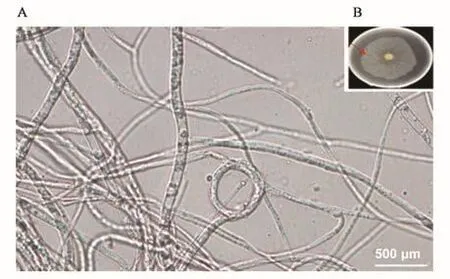
2.1.1 Morphological characteristics of the pathogen From Figure 1, it can be seen that the isolation of the pathogenic bacterial mycelium at first milky white, flat surface, after a period of time the mycelium was velvety, laying growth, can be seen to have a ring of white concentric lines, mycelium dense and the central region tends to be dark yellowish brown. Observed under the microscope mango leaf spot disease pathogen mycelium branching, thin-walled, dark brown, with a diaphragm constriction phenomenon, often entangled in the formation of mycelial ring, did not see the spore structure.

Figure 1 pathogen morphology Fig.1 Morphology of pathogen
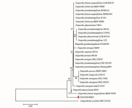
2.1.2 Molecular biological identification of pathogenic bacteria Sequencing results of the ITS amplified fragments and GenBank database (https://www.ncbi.nlm.nih.gov/genbank/) in the sequence comparison, from the results of the comparison of the selection of species closer to the sequence of strains, to NJ method to build a phylogenetic tree, the results show that strain FAFU20190827 was attributed to the genus Diaporthe musigena in the evolutionary tree (Figure 2).

Fig.2 NJ-phylogenetic tree based on ITS sequenceFig.2 NJ-system phylogenetic tree based on ITS sequence
2.2 Characterization of nano magnesium hydroxide
2.2.1 Comparison of the morphology of magnesium hydroxide nanoparticles The size and morphology of magnesium hydroxide nanoparticles were analyzed by XRD and SEM. The results (Fig. 3) showed that the three nano-Mg hydroxide samples synthesized by different methods were consistent with the characteristic peaks of the standard database card (JCPDF-044-1482).The SEM results showed that the synthesized nano-Mg hydroxides showed different morphologies and were more likely to agglomerate into particles. The morphology of nanomagnesium hydroxide synthesized by hydrothermal method at 600 °C was flaky (Fig. 3A), the one synthesized at 80 °C was petal-like (Fig. 3B), and the morphology of nanomagnesium hydroxide synthesized by chemical precipitation method was hexagonal (Fig. 3C). The dimensions of the three types of magnesium hydroxide nanoparticles in the (101) plane were found to be 60.50, 13.53, and 11.62 nm, respectively, according to Scheele’s formula.
2prepared-by-different-methods.webp)
Fig.3 XRD and SEM images of nano magnesium hydroxide prepared by different methodsFig.3 XRD and SEM images of nano-Mg(OH)2prepared by different methods
2.2.2 Specific surface area and zeta potential of nano-magnesium hydroxide As can be seen in Fig. 4, the specific surface areas of MHNPs-MgO600, MHNPs-MgO80 and MHNPs-MgCl2 samples were (14.88±0.10) (92.61±0.52) and (77.42±0.64) m2-g-1 , respectively, which indicated that, using MgO as the raw material for the synthesis of magnesium hydroxide, the high temperature of 600°C is not conducive to the pore formation of magnesium hydroxide nanoparticles, which makes its specific surface area much smaller than that of magnesium hydroxide synthesized at 80°C. The specific surface area of magnesium hydroxide nanoparticles synthesized by choosing magnesium chloride as the raw material is slightly smaller than that of magnesium hydroxide synthesized using magnesium oxide at 80°C. Therefore, different synthesized raw materials and temperatures have an effect on the specific surface area of nanomagnesium hydroxide.The surface zeta potentials of the three types of nanomagnesium hydroxide were (47.28±1.89) (26.65±1.64) and (32.15±1.25) mV, respectively, and the MHNPs-MgO80 had the lowest surface charge and was less stable than the other two types of nanomagnesium hydroxide in aqueous solution. magnesium hydroxide. The nano-magnesium hydroxide prepared by different synthesis methods have large differences in physicochemical properties such as morphology, size and surface area, and the antimicrobial activity of the nanomagnesium hydroxide synthesized by different methods is also directly related to its physicochemical properties.
2prepared-by-different-methods.webp)
Fig.4 BET surface area and Zeta potential of nano magnesium hydroxide prepared by different methodsFig.4 BET surface area and Zeta potential of nano-Mg(OH)2prepared by different methods
2.3 Inhibitory effect of different nano-magnesium hydroxide on mango leaf spot pathogenic fungi
As can be seen from Fig. 5, the growth diameter of the fungus on the plate with the addition of nano-Mg(OH)2 was significantly reduced, indicating that the three kinds of nano-Mg(OH)2 had good inhibition on the growth of mango leaf spot pathogen fungi, and the inhibitory effect was as follows in descending order: MHNPs-MgO80>MHNPs-MgO600>MHNPs-MgCl2, and the higher the mass concentration of nano-Mg(OH)2, the more obvious the inhibitory effect was. effect is more obvious.
2.webp)
Fig.5 Pathogen growth of mango leaf spot after nano hydroxide treatmentFig.5 Pathogen growth of mango phoma leaf spot treated with different nano-Mg(OH)2
All three nano-Mg(OH)2 hydroxides at 25 mg-mL-1 inhibited mycelial growth by more than 40%, while MHNPs-MgO80 inhibited mycelial growth by up to 71.11% at 50 mg-mL-1 (Table 1). The results of the antimicrobial experiments showed that there were large differences in the antimicrobial effects of the three types of nano-magnesium hydroxide, which may be directly related to the above physicochemical properties and so on.
2.webp)
Table 1 Inhibition rate of pathogen of mango phoma leaf spot at different concentrations for three nano-Mg(OH)2
3 Discussion
The use of chemical fungicides to control plant diseases is one of the most important measures to ensure high and stable crop yields, but pathogenic bacteria can easily develop resistance to chemical fungicides, which ultimately leads to the failure of chemical control. Compared with organic and natural antimicrobial agents, inorganic nanomaterials have the advantages of low toxicity, low harm to environmental ecology and human health and high thermal stability as well as pathogenic bacteria are not easy to develop resistance. Nano-magnesium hydroxide has long-lasting and broad-spectrum antimicrobial activity and is a safe, non-toxic, environmentally friendly antimicrobial material with great potential for application. Compared with other nanomaterials, nanomagnesium hydroxide is simple and low-cost to source. Currently, studies on the antibacterial properties of magnesium hydroxide nanoparticles have mainly evaluated the antibacterial activity of single morphology of magnesium hydroxide nanoparticles against model pathogens such as Escherichia coli and Staphylococcus aureus, but studies analyzing the differences in the antibacterial activity of magnesium hydroxide nanoparticles against fruit diseases and the effect of morphology and size on their antibacterial activity have been rarely reported.
In this study, the pathogenic fungus was isolated from mango leaves and identified morphologically and molecularly as the causal agent of mango stem pitting mold leaf spot disease, which mainly affects the leaves, causing spotting and drying and affecting the photosynthesis of mango. As a relatively common disease in mango production, mango leaf spot disease has a great impact on the yield and quality of mango in China. The three kinds of nano-magnesium hydroxide synthesized and prepared in this study have good inhibitory activity against mango leaf spot pathogen fungi, and the higher the mass concentration, the stronger the inhibitory activity, the inhibitory activity of the size of the MHNPs-MgO80>MHNPs-MgO600>MHNPs-MgCl2, which is consistent with the trend of the size of the specific surface area of three kinds of nano-magnesium hydroxide, indicating that the specific surface area is an important factor in the inhibition activity of the nanomagnesium hydroxide antibacterial activity. In addition, through the zeta potential results, it was hypothesized that the reason for the different inhibitory properties of the three types of magnesium hydroxide nanoparticles against the mango leaf spot fungus may be that they have different surface charges, and there are differences in adsorption capacity with the surface of the pathogenic bacteria, and the nanoparticles that are attracted to the surface of the pathogenic bacteria under the interaction of the charge can destroy the integrity of the cell wall of the pathogenic bacteria and increase the permeability of cell membranes, which ultimately This leads to the death of the pathogen. The potential of fungi is generally negative, MHNPs-MgO80 has a smaller surface charge and the largest specific surface area, it is easier to contact with the pathogenic bacteria, so MHNPs-MgO80 has a better antibacterial effect. Studies have shown that the antimicrobial mechanism of nanomaterials mainly includes reactive oxygen species (ROS) generation, metal ion dissolution and contact sterilization, while nano-magnesium hydroxide can damage the cell membrane through contact, resulting in the content flowing out and entering the cell through cytotranspiration, releasing a large amount of OH- in the cell, causing the DNA and protein of the bacterium to be damage and denaturation, thus leading to the death of the bacterium. However, the inhibition mechanism of nano-magnesium hydroxide on the pathogenic fungi of mango leaf spot disease has not been investigated in this study and remains to be explored in depth.
The three different shapes of nano-magnesium hydroxide prepared in this study showed significant inhibitory effects on the pathogenic fungi of mango leaf spot disease, and the differences in the antimicrobial effects were correlated with their own physicochemical properties, such as charge and specific surface area, as well as the mass concentration of the antimicrobial agent. Although the nano-magnesium hydroxide synthesized by different methods all possessed certain antimicrobial properties against the pathogenic fungi, their antimicrobial performances differed significantly, which indicates that the effects of their synthesis methods, physicochemical properties, etc. on the antimicrobial effects should be taken into account when selecting efficient nanoantimicrobial agents. This study provides a new research idea for screening suitable nano antifungal agents, and also provides theoretical guidance for nanomaterials in the prevention and treatment of agricultural pathogens. In the future, we can consider the application of nanoparticles of magnesium hydroxide as an antimicrobial material in the green prevention and control of fruit tree diseases, which on the one hand can reduce the use of traditional chemical pesticides and improve the quality and yield of China’s fruit industry, and on the other hand, can reduce the impact on the environment to protect people’s healthy life.
